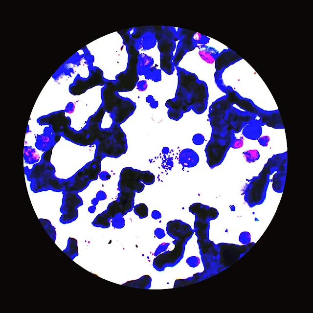
Leishmania donovani bodies refer to the intracellular parasites of the species Leishmania donovani, which are responsible for causing visceral leishmaniasis, also known as kala-azar. Here's an overview of Leishmania donovani bodies:
Morphology:
- Size and Shape: Leishmania donovani bodies are typically small, measuring 2 to 4 micrometers in length. They have an elongated oval or rod-shaped morphology.
- Internal Structures: Within the cytoplasm of the parasite, various structures can be observed, including the nucleus (kinetoplast), flagellum, and other organelles such as the mitochondrion and Golgi apparatus.
Life Cycle:
- Sandfly Stage: The life cycle of Leishmania begins when a female sandfly of the genus Phlebotomus or Lutzomyia ingests blood from an infected mammalian host, thereby acquiring the parasite.
- Promastigote Stage: In the midgut of the sandfly, the ingested Leishmania parasites transform into promastigotes, which are elongated, flagellated forms. These promastigotes multiply and migrate to the anterior midgut and proboscis of the sandfly.
- Transmission to Mammalian Host: During subsequent blood meals, infected sandflies transmit promastigotes to mammalian hosts.
- Amastigote Stage: Upon entering the mammalian host, promastigotes are engulfed by macrophages and transform into amastigotes, which are rounded, non-flagellated forms. These amastigotes multiply within the macrophages and cause infection.
Clinical Significance:
- Leishmania donovani is the causative agent of visceral leishmaniasis (VL), the most severe form of leishmaniasis.
- VL is characterized by fever, weight loss, hepatosplenomegaly (enlargement of the liver and spleen), anaemia, and pancytopenia.
- If left untreated, visceral leishmaniasis can be fatal.
Diagnosis:
- Diagnosis of visceral leishmaniasis is typically made by detecting Leishmania donovani bodies in tissue aspirates, such as bone marrow, spleen, or lymph nodes, using microscopy.
- Molecular techniques, such as polymerase chain reaction (PCR), can also be used for diagnosis, particularly in cases where microscopy may yield false-negative results.
Treatment:
- Treatment of visceral leishmaniasis usually involves antimonial drugs (such as sodium stibogluconate or meglumine antimoniate), amphotericin B, miltefosine, or paromomycin.
- Combination therapy may be used in some cases to enhance efficacy and reduce the risk of drug resistance.
In summary, Leishmania donovani bodies are the intracellular parasites responsible for causing visceral leishmaniasis, a severe and potentially fatal disease. Early diagnosis and appropriate treatment are essential for the management of visceral leishmaniasis.


No comments:
Post a Comment